
When nuclear energy is mentioned, many people immediately think of nuclear power plants or even atomic bombs. In reality, however, nuclear energy is being widely applied in everyday life, particularly in healthcare. In Dong Nai, nuclear energy is gradually moving beyond X-ray and CT (computed tomography) rooms and radiation therapy units into a broader space encompassing research, training, agriculture, environment, industry, logistics and clean energy.
Part 1: The “magic eye” of modern medicine
From tiny, hard-to-detect lesions to complex diseases inside the body, X-rays, CT scans, mammography, bone density measurements and digital subtraction angiography (DSA) are becoming the “magic eyes” of modern medicine.
By producing detailed images, enabling early disease detection and supporting precise interventions, nuclear-energy-based techniques are playing an important role in improving diagnosis and treatment and protecting patients’ health.
Saving lives in time with invisible X-rays
In late 2023, Dong Nai Children’s Hospital admitted P.N.M.T., a four-year-old patient from Long Binh Ward, one day after she fell from a high loft onto the ground. The girl’s mother said she had checked her daughter after the fall but found no unusual signs. Since the child did not complain of pain, she did not take her to the hospital. It was not until the following day, when the girl complained of a headache and began vomiting, that her family took her to the hospital.
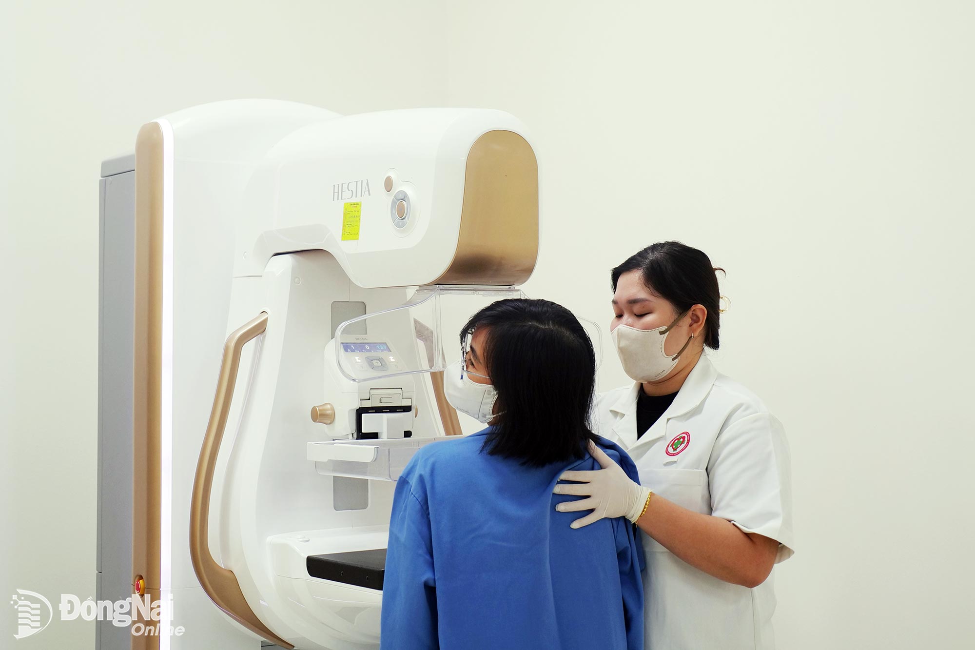 |
| Patients undergo mammography screening for breast cancer at University Medical Shing Mark Hospital. |
After taking the patient’s medical history and suspecting a brain injury, doctors immediately ordered a CT scan. The scan revealed a very large intracranial hematoma, about the size of an orange in the girl’s head, compressing the right side of the brain. The patient underwent emergency surgery to remove the hematoma.
Dr. Nguyen Van Toan, a Level I specialist in neurosurgery, former doctor at Dong Nai Children’s Hospital who directly treated the girl, said this was a typical case of severe brain injury that initially showed no external signs of injury, causing the family to underestimate its seriousness and delay medical attention. He said this was also one of many cases that were diagnosed and treated surgically in time, thereby avoiding the risk of death.
According to Dr. Toan, CT scans help doctors diagnose injuries and conditions such as brain hemorrhage, fractures, depressed skull fractures and diffuse axonal injury, as well as diseases including brain tumors and cerebral vascular malformations. CT scans not only help doctors detect diseases but also enable them to assess disease severity, facilitating the development of appropriate medical or surgical treatment plans.
CT and X-ray imaging are important not only in neurosurgery but also in diagnosing surgical conditions and orthopedic trauma.
Dr. Nguyen Quoc Lu, a Level II specialist and Acting Head of the Department of General Surgery, Orthopedic Trauma and Burns at Thong Nhat General Hospital, said X-rays are usually the first imaging method ordered for trauma cases, particularly when fractures or dislocations are suspected. X-ray images allow doctors to determine the location and degree of displacement of a fracture, assess dislocations and decide on appropriate treatment.
However, not all injuries can be fully assessed using X-rays. For complex trauma, multiple-fragment fractures, fractures involving joints, the spine or pelvis, or injuries that are difficult to see on conventional X-rays, CT scans are highly valuable. CT provides more detailed images and can reconstruct images in multiple planes and even in 3D, enabling doctors to more accurately assess the location and severity of injuries and their relationship with surrounding structures.
In surgery, CT also helps detect and assess many internal injuries, such as trauma to the chest, abdomen and pelvis. This enables doctors to quickly identify injuries and select appropriate treatment methods for each patient, particularly in emergency situations.
“Decoding” the body and screening for cancer
At University Medical Shing Mark Hospital, the nuclear-energy-based diagnostic imaging system currently includes fixed and mobile X-ray machines, a 128-slice CT scanner, a mammography machine, a bone densitometer and a cancer radiation therapy system. Beyond detecting diseases after symptoms appear, radiation technology is expanding opportunities for screening and early diagnosis.
Dr. Luong Dinh Thu, Head of the Department of Diagnostic Imaging at University Medical Shing Mark Hospital, said X-rays are used in mammography to detect abnormalities in breast tissue, including signs suggestive of breast cancer before patients develop obvious symptoms. Meanwhile, dual-energy X-ray absorptiometry (DEXA) can help detect reduced bone density, osteoporosis and the risk of fractures.
“A postmenopausal woman who is diagnosed with osteoporosis at an early stage can receive advice on treatment and fracture prevention. Likewise, a woman whose breast abnormality is detected before symptoms appear has a greater opportunity for early diagnosis and treatment,” Dr. Luong Dinh Thu emphasized.
While X-rays and CT scans allow doctors to observe anatomical structures and morphology, nuclear medicine offers a different approach by focusing on the functions and biological activities of organs.
Dr. Kim Ngoc Si Ha, a Level II specialist and Head of the Department of Oncology and Nuclear Medicine at Dong Nai General Hospital, said SPECT and PET are two nuclear medicine imaging techniques that use small amounts of radiopharmaceuticals to record the functional activity and metabolism of organs in the body. These techniques can help detect bone metastases, assess thyroid disorders, perform renal function scans and evaluate myocardial perfusion, among other applications. The hospital is currently preparing to perform kidney transplants. Renal function scans will help doctors assess the function of each kidney in a patient’s body.
Building a modern technology network
Dr. Nguyen Van Binh, a Level II specialist and Deputy Director of the Department of Health, said nuclear energy in healthcare enables very early diagnosis and the precise destruction of dangerous cancer cells. Through the use of radioactive isotopes and nuclear radiation, nuclear medicine and radiation therapy have transformed tiny physical particles into cutting-edge “weapons” for protecting human health. The application of nuclear energy is a breakthrough technological field that plays a decisive role in transforming and comprehensively improving the quality of medical examination and treatment for residents across the city, he said.
Common applications of nuclear energy in healthcare today include X-rays, CT scans, DSA, mammography, bone densitometry, scintigraphy, SPECT/PET, linear accelerators, radiopharmaceuticals and medical physics.
Since the 2000s, Dong Nai’s healthcare sector has applied nuclear energy by putting several CT scanners into operation at hospitals. About a decade later, Dong Nai General Hospital began using radiation therapy equipment to treat cancer. In 2021, a Halcyon linear accelerator radiation therapy system (Varian, USA) was put into use at University Medical Shing Mark Hospital. These developments have helped improve the capacity to diagnose and treat brain and skull disorders, cancer, stroke and many other complex diseases.
“To date, Dong Nai’s healthcare sector has established a foundation of equipment, human resources and experience in operating advanced medical technologies. This is an important advantage for continuing to expand specialized techniques in the new period, minimizing the need to transfer patients to higher-level facilities, reducing healthcare costs for residents and easing the burden on Central-level hospitals,” Dr. Nguyen Van Binh emphasized.
By Hanh Dung - Translated by Mai Nga, Thu Ha



Thông tin bạn đọc
Đóng Lưu thông tin